 There’s more than meets the eye.
At first glance, few would be able to identify what’s in the images below. But that’s the beauty of microphotography.
The winning photos this year in Nikon’s annual Small World Photomicrography Competition offer an up-close look at our world's microscopic realm -- things that can’t be seen with the naked eye.
Here are some of the winning images.
(Please click on the photos for hi-resolution images)
>> First place
Dr Oscar Ruiz, Houston, Texas, USA

That’s the face of a four-day-old zebrafish embryo. Photograph: Oscar Ruiz/ University of Texas MD Anderson Cancer Center
>> Second place
Douglas L Moore, Stevens Point, Wisconsin, USA

This vibrant specimen captured by Moore is a polished slab of Teepee Canyon Agate - a 273-million-year-old marine sedimentary agate in the Black Hills of western South Dakota. TeepeeCanyon Agate is striking both macroscopically and microscopically, with vibrant colours and wide bands alternating between chalcedony fibers and iron oxide particles boldly displayed in this specimen. Photograph: Douglas L Moore/ University of Wisconsin - Stevens Point
>> Third place
Rebecca Nutbrown, United Kingdom

Culture of neurons (stained green) derived from human skin cells and Schwann cells, a second type of brain cell (stained red). Photograph: Rebecca Nutbrown/University of Oxford, Nuffield Department of Clinical Neurosciences
>> Sixth place
Marek Mis, Poland
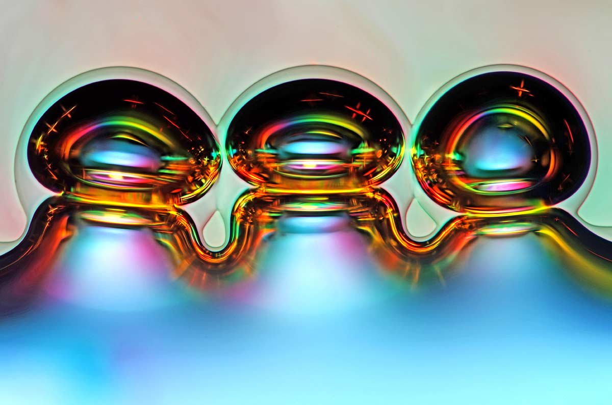
Air bubbles formed from melted ascorbic acid crystals. Photograph: Marek Mis
>> Eleventh place
Francis Sneyers, Belgium

Scales of a butterfly wing underside (Vanessa atalanta). Photograph: Francis Sneyers
>> Thirteenth place
Walter Piorkowski, Illinois

Poison fangs of a centipede (Lithobius erythrocephalus). Photograph: Walter Piorkowski
>> Twentieth place
Michael Crutchley, United Kingdom

That’s cow dung. Photograph: Michael Crutchley
>> Honourable mention
Evan Darling, New York, USA

Scales of a butterfly wing. Photograph: Evan Darling/ Memorial Sloan Kettering Cancer Center
>> Images of Distinction
Erno Endre Gergley, Romania

A Green bottle fly. Photograph: Erno Endre Gergley/Azorean Biodiversity Group
>> Images of Distinction
Matt Inman, Australia

Beta-alanine and taurine crystals. Photograph: Matt Inman
>> Honourable mention
Hei Ming Lai and Dr Wutian Wu, Hong Kong

Dentate gyrus of an optically-cleared transgenic mouse brain in 3D. Photograph: Hei Ming Lai and Dr Wutian Wu/University of Hong Kong
>> >> Honourable mention
Dr Keunyoung Kim, San Diego, USA

Retinal ganglion cells in the whole-mounted mouse retina. Photograph: Dr Keunyoung Kim/University of California, San Diego, National Center for Microscopy and Imaging Research
>> Honourable mention
Jacek Myslowski, Poland
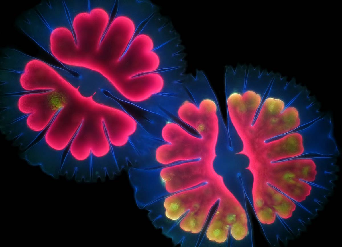
Micrasterias thomasiana (algae). Photograph: Jacek Myslowski
>> Images of Distinction
Dr Talley Lambert, USA

Actin (pink), mitochondria (black), and DNA (red) in a bovine pulmonary artery endothelial cell. Photograph: Dr Talley Lambert/Harvard Medical School
>> Images of Distinction
Yousef Al Habshi, United Arab Emirates

Eyes of a jumping spider (Hasarius adansoni). Photograph: Yousef Al Habshi


 © 2025